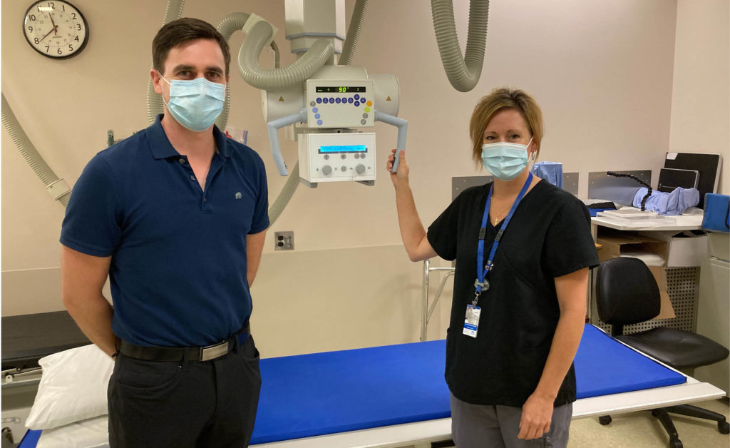
by Dr. David Thomas and Kim Loucks, MRT Medical Imaging, Saanich Peninsula Hospital
Medical imaging touches nearly every disease or condition. At the Saanich Peninsula Hospital (SPH), medical radiological technologists including Kim Loucks and radiologists such as Dr. David Thomas work together to obtain images to help your medical team determine diagnosis and treatment.
Persistent headaches that can’t be explained, pain or trauma caused by a fall or accident, or abdominal pain that doesn’t go away all are reasons your physician may send you for a diagnostic imaging exam. With the remarkable ability to see under your skin and into your organs, diagnostic imaging provides answers to medical questions previously only understood through exploratory surgery.
X-ray and fluoroscopy machines use a small amount of radiation to acquire images, which are commonly used to diagnose fractures, chest congestion and blockages in the gastrointestinal tract and other organs. Advances in X-ray imaging technology have had a significant impact on patient care with improvements in image quality, reduction in radiation dose resulting in better patient diagnosis, and reduced need for exploratory surgery. In 1974, when a small hospital first appeared on Mt. Newton Cross Road, diagnostic imaging options were limited to a basic X-ray, exposed on silver laden film that was developed in a processing method that created fumes and took four minutes to produce a single finished radiograph. Today at SPH you will find a modern medical imaging department that still offers X-ray services, but now also has ultrasound, CT scanning, Bone Mineral Density, and fluoroscopic guided therapeutic injections. Multiple images are acquired digitally, and available immediately to physicians, with your imaging results from the radiologist report also available to you as a patient through Island Health’s MyHealth website.
In addition to X-ray services, SPH’s advanced, multi-slice computerized tomography (CT) scanner is used to detect tumours, infections, internal bleeding and trauma, and blood vessel blockages (CT angiogram). If patients are undergoing treatment for cancer, they can obtain their follow-up CT scan at SPH as well, with vascular access services available to access in-dwelling ports or PICC lines. Ultrasound is used to diagnose abdominal pain and liver disease, detect kidney stones and blockages in the veins and arteries, and guide biopsies.
Over 20,000 X-ray exams are completed at SPH each year. Medical Radiological Technologists are trained to help position patients and, taking the necessary views, the radiologists review and report to a patient’s doctor. X-rays will continue to be used to help physicians diagnose and treat medical conditions.
“It is impressive to see the positive feedback we receive from our patients. Patients can be assured of receiving the highest standards of safety, care and excellence in Medical Imaging,” says Corilee Watters, Manager, Quality, Medical Imaging. “Patients can be confident that the quality of medical imaging available at SPH is equivalent to the best hospitals in Canada.” The SPH Medical Imaging department has received the College of Physicians and Surgeons of British Columbia Diagnostic Accreditation Program Certificate of Accreditation.